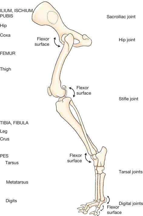
Dog anatomy is not very difficult to understand if a labeled diagram is present to provide a graphic illustration of the same. The stifle or knee is the joint that sits on the front of the hind leg in line with the abdomen.

The back runs from the point of the shoulders to the end of the rib cage.
Anatomy of a dog leg. The upper arm is located below the shoulder. Numerous bone is the long bone of the upper arm which goes all the way to the elbow. The elbow is located below the chest at the back of the foreleg.
This is the first joint in the leg. The forearm is the long bone that runs just after the elbow. Dog leg anatomy is complex especially dog knees which are found on the hind legs.
The technical term for a dog knee is the stifle joint. The stifle joint connects the femur which is the dog thigh bone to the tibia and fibula the lower leg bones and the patellathe canine equivalent to the knee cap. Dogs legs are comprised of bones muscles ligaments and tendons.
The anatomy of a dogs hind leg and foreleg differs just as a human arm and leg differ according to For Dummies. Directly below the shoulder of the foreleg is the humerus bone which ends at the elbow the first joint located just below the chest on the back of the foreleg. Dog Leg Anatomy Just like humans have arms and legs dogs have forelegs and hind legs.
Two thirds of a dogs body weight is carried on their front legs. Only one third is carried on their hind legs. The upper thigh femur is the part of the dogs leg situated above the knee on the hind leg.
The stifle or knee is the joint that sits on the front of the hind leg in line with the abdomen. The lower thigh tibia and fibula is the part of the hind leg beneath the knee to the hock. Dog anatomy comprises the anatomical studies of the visible parts of the body of a domestic dogDetails of structures vary tremendously from breed to breed more than in any other animal species wild or domesticated as dogs are highly variable in height and weight.
The smallest known adult dog was a Yorkshire Terrier that stood only 63 cm 25 in at the shoulder 95 cm 37 in in length. Baring the canine back and chest. The prosternum is the top of the sternum a bone that ties the rib cage together.
The chest is the entire rib cage of the dog. The back runs from the point of the shoulders to the end of the rib cage. The term back is sometimes used to describe the back and the.
Dog anatomy is not very difficult to understand if a labeled diagram is present to provide a graphic illustration of the same. That is exactly what you will find in this DogAppy article. It provides information about a dogs skeletal reproductive internal and external anatomy along with accompanying labeled diagrams.
The dog paw has five basic parts. A the claw B digital pads C metacarpal on the front paws and metatarsal on the rear paws pad D dew claw E carpal pad. The metacarpal metatarsal and digital pads function as the load-bearing shock-absorbing pads.
The carpal pad helps with skid and traction on a slope or while stopping. Understanding the dog leg anatomy is also important as this is an area that is very much prone to injury. Dog leg problems can be classified to further understand how they should be dealt with and treated.
These dog leg injuries include bone fractures bone cracks ligament tears ligament damage cuts bruises and joint pain. The pelvis is where the femoral large leg bone head fits into the hip joint. Dog Leg Bones Diagram - Dogs Hind Limb Muscle Anatomy In Latin The following 40 files are in this category out of 40 total.
Large dogs can handle larger bones like lamb necks lamb shanks beef leg bones whole rabbit whole chickens or chicken carcassesOrangutan dog pig cow tapir and horse. The cruciate ligaments in simple terms are like two pieces of strong elastic that hold the knee together. If a cruciate ligament is damaged the knee becomes wobbly and often very painful.
The most common way for a dog to damage a cruciate ligament is by jumping skidding twisting or turning awkwardly. Comparative anatomy between dogs and humans has been described in other sources. 1-3 We have chosen to use some terms consistently throughout the chapter rather than use equally acceptable synonyms.
The canine forelimb is known also as the thoracic limb and the pectoral limb but we use the term forelimb. Anatomy of the dog - Illustrated atlas This modules of vet-Anatomy provides a basic foundation in animal anatomy for students of veterinary medicine. This veterinary anatomical atlas includes selected labeling structures to help student to understand and discover animal anatomy skeleton bones muscles joints viscera respiratory system cardiovascular system.
Most of the different causes are related to the dogs spinal column spinal cord or the nerves that supply the back legs. They can be divided into broad categories. Injury to the spinal cord or nerves supplying the hind legs.
This is generally the most obvious cause of dog hind leg weakness. There is a broad range of injuries that are possibly associated with the canine front leg. In reality the anatomy of a dogs leg is very complex.
The bones and ligaments can easily be cracked stretched or twisted when impact is applied through running jumping or by virtue of an accident or jolting impact as listed below. Luxating patella cruciate ligament rupture and torn meniscus are things you may see on a daily basis so its critical that you develop a solid understanding of the anatomy of the canine knee from an early stage in your veterinary career.