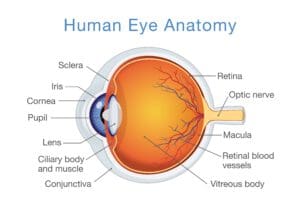
The Optic Nerve the Oculomotor Nerve the Trochlear Nerve and the Abducens Nerve. The eye adopts a position known as down and out.
The trochlear nerve also contributes to the motor innervation of the eye.
Nerves in the eye. Refracts light - bends it as it enters the eye. Controls how much light enters the pupil. Focuses light onto the retina.
Contains the light receptors. Innervation of the eyeball and surrounding structures is provided by the optic oculomotor trochlear abducens and trigeminal cranial nerves. This article covers the anatomy function and clinical relevance of the vessels and nerves of the eye.
Key facts about the neurovasculature of the eye. Four Cranial Nerve pairs control the eyes themselves including. The Optic Nerve the Oculomotor Nerve the Trochlear Nerve and the Abducens Nerve.
Cranial Nerve 2 CN II - Optic Nerve. CNII Cranial Nerve 2 carries Vision to the brain. This nerve does not contain Schwann cells.
The optic nerve also known as cranial nerve II or simply as CN II is a paired cranial nerve that transmits visual information from the retina to the brain. In humans the optic nerve is derived from optic stalks during the seventh week of development and is composed of retinal ganglion cell axons and glial cells. It extends from the optic disc to the optic chiasma and continues as the optic tract to the lateral geniculate nucleus pretectal nuclei and superior colliculus.
The optic nerve is the sensory nerve that involves vision. When light enters your eye it comes into contact with special receptors in your retina called rods and cones. Rods are found in large.
The optic nerve is located in the back of the eye. It is also called the second cranial nerve or cranial nerve II. It is the second of several pairs of cranial nerves.
The job of the optic nerve is. Light passes through the eyeball to the retina. There are two main types of light receptors - rods and cones.
Rods are more sensitive to light than cones so they are useful for seeing in dim light. Optic nerve CN II. Sensory nerve that transmits impulses from the retina to the brain Oculomotor nerve CN III trochlear nerve CN IV and abducent nerve CN VI.
Enter the orbital space through the. Ophthalmic nerve part of the trigeminal nerve CN V. This nerve has three branches.
The optic disc identifies the start of the optic nerve where messages from cone and rod cells leave the eye via nerve fibres to the optic centre of the brain. This area is also known as the blind spot. Leaves the eye at the optic disc and transfers all the visual information to the brain.
The trochlear nerve also contributes to the motor innervation of the eye. Of the extraocular muscles it only innervates the superior oblique muscle. Trigeminal Nerve CN V.
Of the 3 branches of the trigeminal nerve the ophthalmic nerve is involved in sensory innervation of the eye. The optic nerve also known as the cranial nerve II runs from the eyes to the brain. It may become pinched.
When pinching of the occipital nerve or nerves in the neck occurs it is known as Occipital neuralgia. Symptoms of Occipital neuralgia are numbness tingling or pain around the base of the skull. Cranial Nerve 4 This is the trochlear nerve which stimulates one of the eye muscles.
Namely the superior oblique muscle. This nerve can be damaged if you undergo any severe injuries to your head. The inside lining of the eye is covered by special light-sensing cells that are collectively called the retina.
It converts light into electrical impulses. Behind the eye your optic nerve carries. Each eye muscle is stimulated by a specific cranial nerve.
The optic nerve a cranial nerve which carries impulses from the retina to the brain as well as other cranial nerves which transmit impulses to each eye muscle travel through the orbit the bony cavity that surrounds the eyeball. Nerves of the eye The main function of the eye is sight and the nerve that enables sight is the optic nerve CN II. Nerves that innervate the extraocular muscles are called bulbomotors and they are the oculomotor CN III trochlear CN IV and abducens CN VI nerves.
Oculomotor nerve CN III A lesion of the oculomotor nerve affects most of the extraocular muscles. The affected eye is displaced laterally by the lateral rectus and inferiorly by the superior oblique. The eye adopts a position known as down and out.
Trochlear nerve CN IV A lesion of CN IV will paralyse the superior oblique muscle. The front end of the optic nerve is visible at the back of the eye when your doctor or an eye specialist looks through the pupil with an ophthalmoscope. The round front end is just over 15 millimeters in size.
Normally the end of the nerve called the optic disc has a crisp outline and is indented slightly.